Recent Posts
Use Endodontic Microscope in Contemporary Endodontics

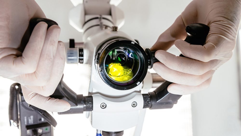
Contemporary endodontics has the advantage of many new technologies that have made root canal treatment more comfortable, faster, and more effective. The dental microscope is a new technology that allows the root canal specialist to see the many details that are vital in endodontic treatment. The attention to detail and precision that is required in endodontic procedures is much helped by the use of these modern dental microscopes.
Because endodontic treatments are generally needed in confined, dark areas of the mouth, it can be challenging to create the kind of precision required for an effective treatment. The outcome of the treatment may come down to mere fractions of millimeters, and the latest technologies are needed to create this precision. This required precision has long required visual aids to help in keeping the procedures precise. It was more than 100 years ago that using microscopes was suggested for endodontic procedures, and the visual aids have gotten more and more refined over time.
Table of Contents
Modern Dental Microscopes
While older dental microscope models gave a small magnification level to aid in precision, the latest standards are higher. In 1998, postgraduate endodontic programs included a microscope proficiency standard. However, the newest standard requires that microscopes use a magnification strength that is stronger than magnifying eyewear. By 2008, 90% of endodontists were using microscopes in their exacting work. The use of these microscopes is now spreading to other dental specialties because of the benefits that endodontists have seen from their use.New Standards, New Skills, New Equipment
As the scientific knowledge behind endodontics has improved over the past decades, so have the technological gains in the field. The improvement of vision that has been provided by the latest endodontic microscopes has allowed for a higher degree of precision that every patient needs for the best outcome. These microscopes both magnify the small areas where the work is being done and provide some illumination to these dark areas.The Benefits of the Latest Design
The loupes worn on the head and attached to glasses in the past had the issue of weight on the head that kept these magnifiers restricted to only the number of lenses most needed for magnification. However, the latest dental microscopes have a very different configuration. These microscopes are created as self-supported units, so there is no need to keep the weight down by restricting the number of lenses. These microscopes can have many lenses and prisms and can provide a much higher degree of magnification because of it. Not only are they better for the visualization of the dental problem, but they are also far more ergonomic to use. The old use of loupes caused eye fatigue and strain because they forced the practitioners’ eyes to rotate because of the angle of the lenses. This medial rotation and the resulting strain meant that the practitioner could suffer eye pain for long procedures or multiple procedures in a day. With the newest dental microscopes, eye strain and fatigue are gone, and the practitioner can continue with challenging procedures. The arrangement of the latest dental microscopes is a parallel orientation of binoculars that is aided by prisms that let light reach the eyes in straight lines. This allows for the easier visualization of distant objects, and it will enable the practitioner to look straight ahead to reduce the eye fatigue and muscle tension caused by the rotation of the eyes needed in older technologies. The body posture required to use the latest microscopes also helps the practitioner to avoid the back and neck pain that was caused by earlier devices.Increased Magnification of Root Canal
The loupes, once pervasive in endodontics, provided a fixed magnification range from 2.5 to 6x magnification. The microscopes now commercially available have adjustable ranges from 4 to 25x magnification. The range of magnification can be changed from low to medium to high magnification depending on the procedure or even the step of the procedure that is being performed. These settings are useful for endodontic work that is both surgical and non-surgical. The low magnification settings, 2 to 8x, are often used for taking an overview of the area being operated on. The middle range of magnification, 8 to 16x, is generally used for root canal work and endodontic surgeries. The highest magnification range, 16 to 25x, is used to identify the tiny structures and fine details of the operating field. The use of these high-powered microscopes makes practitioners more accurate, keeps them more comfortable, and ensures that there will be no fine details missed.Nonsurgical Root Canal
For endodontists, a dental microscope can be used for many clinical procedures- not just for root canal surgeries. These units help diagnose dental problems such as cavities, assessing fracture lines, and identifying when restorative or crown filling margins are insufficient. Microscopes can help with the treatment of internal resorption and help the practitioner see when the dentin color or texture has changed. Being able to see everything in such detail can help during virtually every phase of endodontic treatment. It can help remove dentin more safely, assist in the treatment of calcified canals, and the removal of calcifications and denticles in canals. It is also helpful in post-operative procedures such as the treatment of fused teeth and finding and removing leftover materials such as pastes and leftovers from fillings. These microscopes are also used to repair perforations and to clean the site afterward. Modern dental microscopes have mainly transformed contemporary endodontics. This new standard of care has led to endodontic microsurgeries that are more precise thanks to the ability of the practitioner to see the smallest features on the teeth and surrounding tissue. Related articles:- >Regenerative Endodontic as an Alternative to Root Canal
- Five things you need to know before a root canal treatment
- Use of CT scan in modern Endodontics
- Best Practice in Contemporary Endodontics


