Affordable Dental Implants in Houston

Addressing Dental Implant Cost Concerns
Are you hesitant to replace missing teeth because you’re worried about the cost of dental implants in Houston? You’re not alone. Many people put off getting dental implants due to concerns about affordability and quality. It’s true that dental implants have a reputation for being a higher-priced dental procedure – after all, they involve surgical placement and custom-crafted new teeth. However, that doesn’t mean you have to break the bank for a healthy, beautiful smile. Omega Dental in Midtown Houston is committed to providing affordable dental implants Houston patients can trust for top quality and lasting results. We understand the common worries: Will cheaper implants be lower in quality? Is it better to choose dentures because implants cost more? In this introductory section, let’s tackle those concerns head-on.
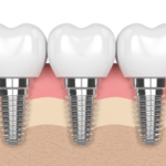
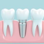
Dental Implants = Long-Term Value: First, it helps to know why implants tend to cost more than other options. A dental implant isn’t just a fake tooth—it’s a permanent replacement for your entire tooth structure. An implant includes a titanium post inserted into your jaw (acting like a tooth root) and a lifelike crown on top. This process requires skilled specialists, advanced technology, and multiple steps to ensure everything is done safely and correctly. While the upfront dental implants cost is higher than, say, a denture or bridge, implants can last decades or even a lifetime with proper care. In contrast, dentures often need replacement every 5–10 years. So even though you pay more now, you save money and hassle in the long run because you won’t need frequent replacements (Dental Implant Cost & Care | Omega Dental Houston TX) (All-on-4 Dental Implants in Houston Explained | Omega Dental Houston TX). Patients often find that the confidence and comfort implants provide – being able to eat, laugh, and smile naturally again – are well worth the investment.
Quality vs. Affordability – You Can Have Both: We’ve all heard the phrase “you get what you pay for.” When it comes to dental work, you certainly don’t want bargain-basement implants from an unqualified provider. But affordable doesn’t have to mean low quality. Omega Dental aims to bridge the gap between cost and quality. Our clinic offers competitive pricing for implants without cutting corners on materials or expertise. Every implant case at Omega Dental is handled by experienced specialists using top-grade components (like titanium implants and porcelain crowns) that meet strict FDA standards. We simply believe that fair pricing and excellent care should go hand in hand. In fact, one of our core missions is making high-quality dental implants accessible to more Houston residents. If you’ve been searching online for “affordable dental implants Houston” and feeling skeptical, rest assured – at Omega Dental, you truly can get premium implant treatment at a fair, manageable cost.
What Do Dental Implants Cost in Houston? It’s natural to want real numbers. While every patient’s needs are unique, here are some typical price ranges for dental implants in Houston to give you an idea:
- Single Tooth Implant: Approximately $3,000 – $6,000 for one implant, abutment, and crown (Dental Implant Cost & Care | Omega Dental Houston TX). This covers replacing one missing tooth with a complete implant restoration.
- Multiple Teeth Implants: Roughly $6,000 – $10,000 when replacing two or three teeth with implants (Dental Implant Cost & Care | Omega Dental Houston TX). The cost goes up with more teeth or if additional procedures (like bone grafts) are needed.
- Full Mouth Implants: Anywhere from $20,000 up to $40,000 (or more) to replace all teeth on one or both arches with implant-supported teeth (Dental Implant Cost & Care | Omega Dental Houston TX). This could involve advanced techniques like All-on-4 implant bridges for a whole new set of teeth in one jaw.
These ranges might seem broad, but that’s because the cost of dental implants in Houston truly depends on individual factors – how many implants, your oral health, the technology used, etc. A straightforward single implant is at the lower end, while a complex full-mouth reconstruction is at the higher end. The good news is that Omega Dental’s fees are highly competitive within these ranges. We strive to keep our prices fair for the Houston community. In fact, for sophisticated full-arch implant treatments like the All-on-4 “Teeth in a Day”, Omega Dental is known for affordable pricing – about $25,000 per arch on average at our office, which often beats other providers. Our goal is to maximize value for our patients: you get exceptional outcomes and a reasonable price.
So, if sticker shock has been holding you back from even exploring implants, take heart. Omega Dental is here to make it work for you. Next, we’ll discuss how we make implants more affordable – from free consultations to flexible financing and promotions – without sacrificing the world-class quality and expertise you deserve.
Cost-Effective Solutions and Promotions for Patients
Worried about the price tag of implants? Omega Dental has a variety of patient-friendly services and promotions to ensure dental implants cost is manageable. We firmly believe that everyone deserves a healthy smile, so we go the extra mile to remove financial barriers. Here’s how we make your treatment budget-friendly:
1. Free Implant Consultations: We offer free consultations for new patients interested in dental implants. This means you can meet with our implant experts, get an evaluation (often including necessary X-rays or 3D scans), and discuss your treatment options at no cost. Many dental offices charge $150–$300 just for an implant consultation (What is The Cost of a Single Tooth Implant? | Omega Dental), but at Omega Dental your initial visit is complimentary. This free consult is a zero-pressure opportunity for you to learn about the process and get a personalized quote. We’ll explain the procedure, answer all your questions, and give you a clear breakdown of the fees involved. Patients really appreciate this transparency and the chance to get professional advice for free. It’s part of our commitment to earning your trust from day one. (As a bonus, we often provide a free panoramic X-ray or CT scan during the consultation if needed, which helps us assess your case thoroughly.) Why do we do this? We want you to feel comfortable and informed about investing in your smile, without worrying about an initial office charge. By removing that hurdle, you can focus on deciding what’s best for your health.
2. Same-Day Dental Implant Consultation: If you’re eager to get started quickly, we’ve got you covered here too. Omega Dental offers convenient scheduling – often even same-day dental implant consultations. This means that if you call us in the morning, there’s a good chance we can see you that very day to discuss implants. We keep appointment slots available for new patients and urgent cases because we know tooth loss can sometimes be an immediate concern. Our Midtown Houston location has extended hours (we’re even open on Saturdays), making it easier to find a time that works for you. With our large team of specialists, we can accommodate consults sooner than many offices. Don’t spend weeks on a waiting list – we strive to meet your needs as soon as possible. Quick access to care is part of our patient-first philosophy. If you have an upcoming event or just don’t want to wait to get the ball rolling, ask about a same-day consultation when you contact us. We’ll do our best to make it happen so you can start your journey to a new smile without delay.
3. Special Offers and Promotions: To further help with the cost, Omega Dental frequently runs special promotions for new patients and specific treatments. For example, we have offered free X-rays and exams with certain procedures, discounted pricing on implant packages, or low monthly payment deals. Be sure to check our current offers page for the latest promotions. One of our popular promotions makes implants more accessible by breaking down the cost: some patients qualify for implant payments as low as under $100 per month with approved financing. Imagine restoring your smile for about the cost of a cell phone bill! Our specials change periodically, but they’re all aimed at giving you the best value. Whether it’s a seasonal discount or a referral bonus (we even have a patient referral program: “Give $50, Get $50” credits), we love finding ways to make our patients smile in more ways than one. Affordable care is a cornerstone of our practice, so keep an eye out for new deals.
4. Flexible Financing & Payment Plans: Perhaps the biggest help in affordability comes from our third-party financing and in-house payment plans. Omega Dental partners with reputable healthcare financing companies to offer zero or low-interest payment plans. We work with options like CareCredit® and LendingClub® among others () (All-on-4 Dental Implants in Houston Explained | Omega Dental Houston TX). These allow you to pay for your dental implants in small monthly installments rather than one lump sum. During your visit, our treatment coordinator will walk you through the financing application – it’s quick and straightforward, often giving an instant decision. Many patients are relieved to learn they can split their cost over 12, 18, or even 24 months with little to no interest (on approved credit). For instance, instead of paying $6,000 upfront, you might pay around $250 a month over two years. That’s a game-changer! We also offer in-house payment plans for certain cases, which can be tailored to your budget. Omega Dental never wants cost to prevent you from getting the care you need. By offering financing, we make premium implants attainable on practically any budget. Our team will help you find the best payment solution – we’re pretty creative at finding ways to say “Yes, we can make this work for you.”
5. Maximizing Insurance Benefits: While many dental insurance plans don’t cover implants fully (since implants can be considered a “major” or even elective procedure), our insurance specialists do everything possible to utilize any coverage you do have. Some plans will cover portions of the treatment, like the crown or extractions, or implants if they are deemed medically necessary. We will verify your insurance and handle the paperwork to get you the maximum reimbursement available. Even if insurance only covers a little, we think every bit helps. We’re also happy to provide documentation if you have an HSA/FSA (Health Savings or Flexible Spending Account) so you can use tax-advantaged funds for the procedure. Our office accepts all major PPO dental plans and some Medicare Advantage plans that include dental. Rest assured, we leave no stone unturned in finding ways to offset the cost on your behalf.
In summary, Omega Dental makes paying for dental implants as painless as the procedure itself (which, by the way, is usually very comfortable!). With free consults, prompt appointments, special discounts, and financing plans, we truly live up to our promise of affordable dental care. Next, let’s look at why thousands of patients have trusted us – from our 5-star reviews to advanced technology – and how we’ve become a top choice for dental implants in Houston.
Proven Results: 5-Star Reviews and Advanced Technology
Choosing a dental provider is a big decision. How do you know you’re in the right hands? One way is to look at social proof – the experiences of other patients and the reputation of the clinic. At Omega Dental, we’re proud to have earned the trust of so many patients over the years. Our commitment to excellent outcomes has resulted in hundreds of 5-star reviews and testimonials from people we’ve treated. We are one of Houston’s top-rated dental implant providers, and nothing makes us happier than seeing another patient leave our office with a bright smile and a 5-star experience.
When you read our reviews or talk to our patients, a few themes come up again and again. People comment on the compassionate, personalized care they received – how our team went above and beyond to make them comfortable during a big procedure. They talk about the life-changing results of their implants – being able to eat their favorite foods again or smile confidently in photos. And yes, many mention how they initially feared they couldn’t afford implants, but Omega Dental’s team worked with them to create a payment plan or financing that fit their budget. These success stories and positive reviews are a testament to our core values: affordability, quality, and treating patients like family. We’re grateful that over 20,000 happy patients have trusted Omega Dental with their smiles, and their feedback motivates us to keep raising the bar.
Another pillar of our authority is the use of advanced technology in our clinic. Modern dental implants are a marvel of science, and we’ve invested heavily in state-of-the-art tech to ensure optimal results. For example, Omega Dental uses 3D CT scans (cone beam CT imaging) for precise diagnostics and treatment planning. This advanced imaging allows our specialists to assess your jawbone in three dimensions, identifying the perfect implant placement while avoiding vital structures. We also utilize computer-guided implant surgery tools. Think of this like GPS for implant placement – we create a digital 3D model of your mouth and plan exactly where each implant should go for maximum stability. During surgery, a custom guide helps the doctor place the implant in the pre-planned position with pinpoint accuracy. The result? Shorter procedure times and a perfectly placed implant, which means a stronger, more comfortable outcome for you.
Our on-site technology doesn’t stop there. We have an in-house digital dental lab capable of crafting crowns, bridges, and even full arches of new teeth with incredible precision. Using CAD/CAM milling and 3D printing technology, we can often have your new tooth (or teeth) ready faster and with fewer appointments. For complex cases like All-on-4 implants, our advanced lab and technology enable us to deliver a same-day smile – you come in for your procedure and leave that same day with a beautiful set of fixed provisional teeth. This is a life-changing service we are thrilled to offer in-house. Only a handful of clinics have the expertise and equipment to do “Teeth in a Day” truly in one location – Omega Dental is proud to be one of them.
In short, Omega Dental combines experience and innovation. We stay on the cutting edge of implant dentistry so that our patients benefit from the best that modern dental medicine has to offer. Our skilled team has placed thousands of implants, and with tools like 3D imaging and computer guidance, you can be confident your smile is being restored with expert precision. This blend of personal care, a proven track record (just read our 5-star reviews!), and advanced tech establishes Omega Dental as a trusted authority in implant dentistry.
Why Choose Omega Dental in Midtown Houston?
Houston is a big city, and there are certainly other places to get dental implants. So, what makes Omega Dental stand out? In a nutshell, we offer a unique combination of convenience, comprehensive care, and a patient-centered approach that’s hard to find elsewhere. Here are some of the top reasons patients choose Omega Dental as their dental home:
All-in-One Multi-Specialty Clinic: Omega Dental is a multi-specialty dental clinic, which means we house multiple types of dental specialists under one roof. Instead of referring you all over town, we can handle every step of your dental implant process right here in our Midtown Houston office. Our team includes experienced oral surgeons, periodontists, prosthodontists, endodontists, and orthodontists. Essentially, no matter what dental issue or procedure you might need – from a tricky tooth extraction or gum surgery to the final implant crown – we have the in-house expertise to take care of it. For implants specifically, our on-staff oral surgeons and periodontists (gum and implant specialists) perform the surgical placement, and our prosthodontist or restorative dentists create the beautiful teeth that attach on top. This collaborative approach ensures seamless care. You won’t be shuttled between different offices or have to find an outside specialist for part of your treatment. At Omega, we coordinate everything for you, making the experience more convenient and less stressful. Patients love knowing that the same trusted team will be with them from start to finish.
Convenient Location in Midtown: Located centrally at 106 W Gray St, Houston, TX, Omega Dental is easy to reach for patients across the Houston metro. We’re right in Midtown, bordering Montrose and just minutes from Downtown. That means whether you live in central Houston or are coming from suburbs like Bellaire or the Heights, our office is accessible via major roads. We also have private parking available. Our Midtown spot was chosen to be convenient for busy professionals and families – you could pop in from work downtown or while running errands in the city. Many of our patients appreciate that they don’t have to drive far out of town for specialist care; we’re essentially in the heart of Houston. Being centrally located, close to public transit, and in a safe, bustling area also gives patients peace of mind when visiting. We’re your neighborhood dental specialists, which makes keeping up with follow-ups or additional appointments much easier.
Modern, Comfortable Facility: When you step into Omega Dental, you’ll notice it doesn’t feel like a typical clinic – it’s modern, welcoming, and designed for patient comfort. We have a spacious, light-filled reception area, and our treatment rooms are equipped with the latest dental chairs and monitoring equipment. We even provide cozy touches like blankets, pillows, and noise-cancelling headphones to help you relax during longer procedures. Our investment in a state-of-the-art facility isn’t just about looks; it’s about creating a safe and comfortable environment for advanced dental care. From strict sterilization protocols to having emergency backup power and monitoring systems, we take your safety seriously. Plus, the ambiance is more spa-like than clinical, which helps ease any anxiety. Patients often comment that our office doesn’t “feel like a dentist” – and we consider that a big compliment!
Multilingual Staff (English & Spanish): Houston is a wonderfully diverse city, and our team at Omega Dental reflects that diversity. We have a multilingual staff, with many team members fluent in Spanish (as well as other languages in some cases). This is a huge benefit if English isn’t your first language or you feel more comfortable discussing medical information in Spanish. From the front desk to the dental assistants and doctors, we can accommodate Spanish-speaking patients so that nothing gets lost in translation. We’ll explain your treatment, finances, and instructions in the language you’re most comfortable with. Communication is key in healthcare, and we never want language to be a barrier. Se habla Español – estamos aquí para responder todas tus preguntas y hacer tu experiencia lo más cómoda posible. By being attentive to language and cultural needs, we make sure all our patients feel welcome and understood. Our goal is for you to feel at home at Omega Dental, regardless of your background.
Personalized Care and Integrity: Lastly – and perhaps most importantly – Omega Dental genuinely cares about you as a person, not just a patient. We know that dental visits, especially for something like implants or surgery, can be intimidating. Our team is known for its friendly, compassionate approach. We listen to your concerns, thoroughly explain your options, and treat you with respect at every step. You’ll never feel like a number here. We schedule ample time for each appointment so things never feel rushed. If you’re nervous, we offer sedation options to ensure your comfort. If you have questions, we’re always ready with answers – no question is too small. This empathetic approach is part of our culture. Moreover, integrity guides everything we do. We will never recommend unnecessary treatments or surprise you with hidden costs. What we will do is give you honest advice and work with you on a treatment plan that fits your needs and budget. Our long-term patients know that Omega Dental is a place they can trust with their family’s smiles year after year.
In summary, Omega Dental offers an unmatched combination of comprehensive specialty care, convenience, and compassion. Our Midtown location is easy to access. Our team of in-house specialists means one-stop care. Our advanced tech means top-notch results. Our friendly bilingual staff makes every visit comfortable. And our patient-centric philosophy means you’ll be treated like family. These are just some of the reasons why so many patients choose Omega Dental and happily refer their friends and relatives to us. We truly love what we do, and it shows in the smiles we create!
Ready for Your New Smile? Book a Free Consultation Today!
If you’ve been dreaming of a confident smile and are interested in dental implants, now is the time to take the next step. We invite you to experience the Omega Dental difference for yourself. Take advantage of our free implant consultation offer and see how affordable and life-changing dental implants can be. Getting started is easy: simply book your appointment online or give our friendly front desk a call. You can request your consultation through our website or by phone, and we’ll find a convenient time that fits your schedule (remember, we often have availability for same-day consultations!).
During your visit, our team will perform a thorough examination and discuss what dental implant solution is best for you – whether you need a single tooth replaced or a full-mouth restoration. You’ll receive a clear treatment plan, including exact costs and timelines, so you can make an informed decision. And of course, this initial visit is completely free and with no obligation. We’re confident that once you meet our team and see our office, you’ll feel reassured that Omega Dental is the right place for your smile journey.
Imagine yourself a few months from now: eating, speaking, and smiling with total confidence thanks to beautiful new dental implants. That outcome is within reach, and it all starts with a simple consultation. We’re here to guide you every step of the way – from consultation to completion – ensuring your experience is smooth and rewarding. Don’t let concerns about cost or fear of the unknown hold you back from the smile you deserve. As we’ve outlined, we have numerous financing and support options to make your implant treatment feasible. Our patients often tell us their only regret is not coming to see us sooner!
Your path to a healthier, happier smile is just one phone call or click away. Schedule your free dental implant consultation with Omega Dental Specialists in Houston today, and let us show you how easy and affordable restoring your smile can be. You can also visit our Dental Implants Services page for more information about the implant solutions we offer and see before-and-after photos of our work. We’re excited to welcome you to the Omega Dental family and be a part of your smile transformation.
Don’t wait – contact Omega Dental now to book your free consultation and take the first step toward a new smile and a new you. We’ve helped over twenty thousand patients reclaim their oral health and confidence, and we’d be honored to help you do the same. Let us give you a reason to smile again!
Omega Dental – Houston’s Home for Affordable, High-Quality Dental Implants. Schedule your consultation at OmegaDentists.com or call us at 713-322-7474. We can’t wait to help you love your smile!


